

Health Information


Learn about different types of bone fractures

Learn about different types of bone fractures (by Sportsperformance Physiotherapy)
What is a fracture?
A fracture occurs when a bone breaks or cracks due to a force that exceeds the bone's ability to flex and bend. Typically, this injury causes severe pain and reduced mobility near the spot of the damage. Fractures can come about in various ways but fall, sports accidents, crashes, fragile bones with age, and overuse injuries are the causes.
The kind of fracture that develops depends on the magnitude of force applied to the bone and the angle of that force. Different fracture types can result from diverse causes. For example, a bone break from a sports injury or motor vehicle accident may produce another fracture compared to one arising from osteoporosis or the repetitive stress of certain athletic activities like running or jumping. High-impact events with one massive force tend to cause more complete breaks, cracks or splintering of the bone.

Different types of fractures:
-
Closed (simple) fracture: The fractured bone has not broken through the skin.
-
Open (compound) fracture: The broken bone may have pierced the skin, or a surface wound may have broken the skin over the site of the bone break.
-
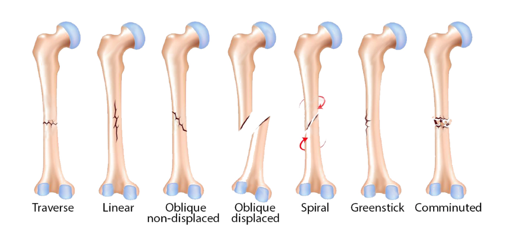
Non-displaced fractures: the broken ends align versus oblique fractures where the break angle is skewed.
-
Oblique fracture: The fracture has an angled pattern and can either be non-displaced (the ends are aligned) or displaced (the ends are not aligned).
-
Transverse fractures: involves a horizontal break, while comminuted fractures shatter the bone into three or more pieces, often from severe impact.
-
Comminuted fracture: The bone has shattered into three or more pieces (usually due to an extreme force or crush-type injury).
-
Greenstick fractures: seen mainly in children, involve an incomplete break through one cortex due to their bendable bones.
-
Compression fractures (e.g. stress fractures): often impact vertebrae in older people with osteoporosis, where two bones are compressed against each other.
-
Hairline fractures (e.g. stress fractures) involve thin cracks from repetitive movement, like stress fractures in runners. Athletes seek physical therapy to rehab hairline fractures.
Fractures can impact any bone in the body. Among the most common injuries orthopaedic surgeons treat are fractures of the toes, feet, lower legs like the shin and femur, and upper limbs, especially the forearm, shoulders, wrists, ribs, vertebrae and hips. Hip fractures are a particular concern in senior citizens because they can result in severe impairment and loss of independence.
How to know if I got a bone fracture?
Several types of medical imaging tests can help identify and characterise bone fractures, ensuring accurate diagnosis and proper management. One or more of the following exams may be ordered:
-
X-rays represent a first-line test to confirm fractures and assess their displacement, severity and complications. However, simple x-rays cannot identify all types of fractures.
-
Magnetic resonance imaging (also known as “MRI”) utilizes magnets and radio waves to generate 3D images of bones and surrounding structures like cartilage, ligaments and soft tissues. An MRI can detect subtle fractures missed on x-rays and elucidate the full extent of damage.
-
CT scans provide more detailed, cross-sectional views of bones compared to x-rays and can precisely depict fracture patterns to guide surgery if needed.
-
Bone scan may be ordered if x-rays fail to detect a suspected fracture. The scan involves injecting a small amount of radioactive material and capturing its accumulation at sites of injury over several hours. Bone scans can locate stress fractures and other covert breaks.
Your healthcare provider will determine the best initial and follow-up imaging based on your symptoms, physical exam findings and condition. No single test definitively diagnosed all fracture types so a combination may be used based on clinical suspicion to rule out other mimickers of bone injury. Imaging also tracks fracture healing to ensure adequate progress and detect complications.


